http://www.nlm.nih.gov/medlineplus/ency/images/ency/fullsize/19221.jpg
Structure of the Small Intestine
- Muscular tube extending from the pyloric sphincter to the ileocecal valve
- Longest section of the alimentary tube, averaging 6-13 feet in a living person
- Hangs like sausage-like coils in the abdominal cavity and hangs from the posterior abdominal wall by the fan-shaped mesentary
- Has three subdivisions:
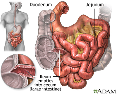
- Duodenum: It curves around the head of the pancreas and is about 10 inches long.
- Jejunum: Extends from the duodenum to the ileum and it about 8 feet long.
- Ileum: About 12 feet long, it is the terminal part of the small intestine. This subdivision joins the large intestine at the ileocecal valve.
Inside the Small Intestine
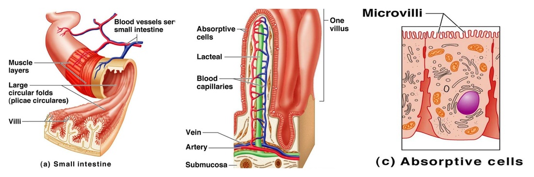
- Microvilli: Tiny projections of the plasma membrane of the mucosa cells that give the cell surface a fuzzy appearance, sometimes referred to as brush border.
- Villi: Fingerlike projections of the mucosa that give a velvety appearance and feel.
- Lacteal: In the villus. A rich capillary bed and a modified lymphatic capillary.
- Circular Folds: Also called plicae circulares, are deep folds of both muscosa and submucosa layers.
- Peyer's Patches: Local collections of lymphatic found in the submucosa.
- Wall is composed of four layers: tunica mucosa (innermost) , tela submucosa, tunica muscularis, and the tunica serosa (outermost)
